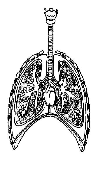
43 diagram of the lungs with labels
Labeled Diagram Of An Labeled diagram of the lungs/respiratory system. 1 (517) 884 6434 email: This may be useful as a printable poster for the classroom, … Blank animal cell diagram worksheet. The volume of a nucleus was considered to be negligible compared to the … There is a similar valve on the left side of the heart, located between the left ventricle and aorta. Lung Diagram Labeled | EdrawMax Template in the following lung labeled diagram, we have shown thyroid cartilage, cricoid cartilage, tracheal cartilage, apex, left upper lobe, hilum, left bronchus, oblique fissure, bronchioles, left lower lobe, base of lung, cardiac notch, right lower lobe, oblique fissure, right middle lobe, horizontal fissure, right bronchus, right upper lobe, and …
Pulmonary Vein: Anatomy, Function, and Significance The four pulmonary veins play an important role in the pulmonary circulation by receiving oxygenated blood from the lungs and delivering it to the left atrium, where it can then enter the left ventricle to be circulated throughout the body. The pulmonary vein is unique in that it is the only vein that carries oxygenated blood. 1.

Diagram of the lungs with labels
30 Fun And Interesting Respiratory System Facts For Kids Lungs. Lungs are a pair of pyramid-shaped organs inside your chest. The primary function of the lungs is to add oxygen to the blood and remove carbon dioxide. This process is called gas exchange or respiration. The various parts of the lungs are: Bronchioles: Bronchioles deliver the air to the gas exchanging unit called alveoli. Bronchioles ... Normal CT chest lung on axial images with labels | e-Anatomy - e-Anatomy Normal anatomy of the thorax on labeled chest CT with scrollable images: radiological anatomy in axial slice of the lungs, mediastinal lymph nodes, trachea, bronchi, pleural cavity, heart and pulmonary vessels. FREE Human Body Systems Labeling with Answer Sheets The free respiratory system labeling sheet includes a blank diagram to fill in the trachea, bronchi, lungs, and larynx. The free nervous system labeling sheet includes blanks to label parts of the brain, spinal cord, ganglion, and nerves. The free muscular system labeling sheet includes a blank diagram to label some of the main muscles in the body.
Diagram of the lungs with labels. 40 human respiratory system with labels Browse 154 diagram of the respiratory system with labels stock photos and images available, or start a new search to explore more stock photos and images. Newest results The respiratory system Lungs with Alveoli Labeled The digestive system lung. Anatomical Position and Directional Terms: Definitions, Example Labeled ... Anatomical Directional Terms: Labeled diagram showing inferior, defined as below or away from the head. Superior (Cranial) and Inferior (Caudal) Superior and inferior also go by different names. Another name for superior is cranial, which makes sense because we are moving toward the cranium or head. Another name for inferior is caudal. Circulatory System Diagram - New Health Advisor There are different types of circulatory system diagrams; some have labels while others don't. The color blue stands for deoxygenated blood while red stands for blood which is oxygenated. Below you'll see diagram specified to the heart, as well as circulatory system diagram of the whole body: How Does the Human Circulatory System Work? 1. Heart How the Lungs Work - The Lungs | NHLBI, NIH The Lungs. Your lungs are the pair of spongy, pinkish-gray organs in your chest. When you inhale (breathe in), air enters your lungs, and oxygen from that air moves to your blood. At the same time, carbon dioxide, a waste gas, moves from your blood to the lungs and is exhaled (breathed out). This process, called gas exchange, is essential to life.
Lung - Pathology Outlines - Histology Lung nontumor - Normal histology. Bronchus has cartilage and bronchial glands, while bronchiole lacks them (Mills: Histology for Pathologists, 5th Edition, 2019) Bronchi and bronchioles up to terminal bronchioles are pure conducting airways, while respiratory bronchioles and alveoli play a role in gas exchange Respiratory system quizzes and labeled diagrams | Kenhub Take a look at the labeled diagram of the respiratory system above. As you can see, there are several structures to learn. Spend a few minutes reviewing the name and location of each one, then try testing your knowledge by filling in your own diagram of the respiratory system (unlabeled) using the PDF download below. Respiratory system unlabeled Anatomy, Thorax, Lungs - StatPearls - NCBI Bookshelf The left lung consists of two lobes: the left upper lobe (LUL) and the left lower lobe (LLL). The right lobe is divided by an oblique and horizontal fissure, where the horizontal fissure divides the upper and middle lobe, and the oblique fissure divides the middle and lower lobes. Fetal Circulation Diagram | Fetal Blood Flow & Circulatory System ... For one, before a baby is born the placenta is the sole source of oxygen whereas in a newborn baby, the lungs are the sole site of oxygen exchange. ... Labeled Diagram of Organs
Body Cavities and Organs with Labeled Diagram - The Major and Minor ... The heart, lung, oesophagus, trachea, bronchi, aorta, brachiocephalic trunk, cranial and caudal vena cava, and vagus nerve. Minor body cavities (in thoracic region) In the thoracic region of an animal body, you will find some minor cavities like the pericardial and pleural cavity. Body Cavities and Membranes: Labeled Diagram, Definitions The 3 meningeal layers are labeled with the stars. The outermost layer of the meninges is the dura mater, which is located beneath the skull. Below the dura mater is the arachnoid, which is the middle meningeal layer. There is a space below the arachnoid called the subarachnoid space, and this is where the CSF is located. Free Respiratory System Worksheets and Printables We have created the Human Body Systems Labeling and Diagramming Worksheet as an instant download for your children. This respiratory system packet includes a fill in the blank diagram to fill in the trachea, bronchi, lungs, and larynx. Respiratory System Diagram - Download this free color diagram of the respiratory system for your kids. The 7 parts of the lungs (and their functions) - LORECENTRAL What are the 7 parts that make up the lungs? 1. Trachea 2. Lobes 3. Bronchi 4. Bronchioles 5. Alveoli 6. Pleura 7. Diaphragm How can I keep my lungs healthy? 1. No smoking 2. Avoid contamination 3. Perform physical exercise 4. Monitor your diet 5. Do not breathe through your mouth Bibliographic references

Habits of the Heart: Lessons: Lung Diagram
Diagram of Human Heart and Blood Circulation in It Ventricle contracts and pushes the blood into the pulmonary artery that sends blood to your lungs from where oxygen-rich blood returns to the left ventricle and the process continues. Exterior of the Human Heart A heart diagram labeled will provide plenty of information about the structure of your heart, including the wall of your heart.
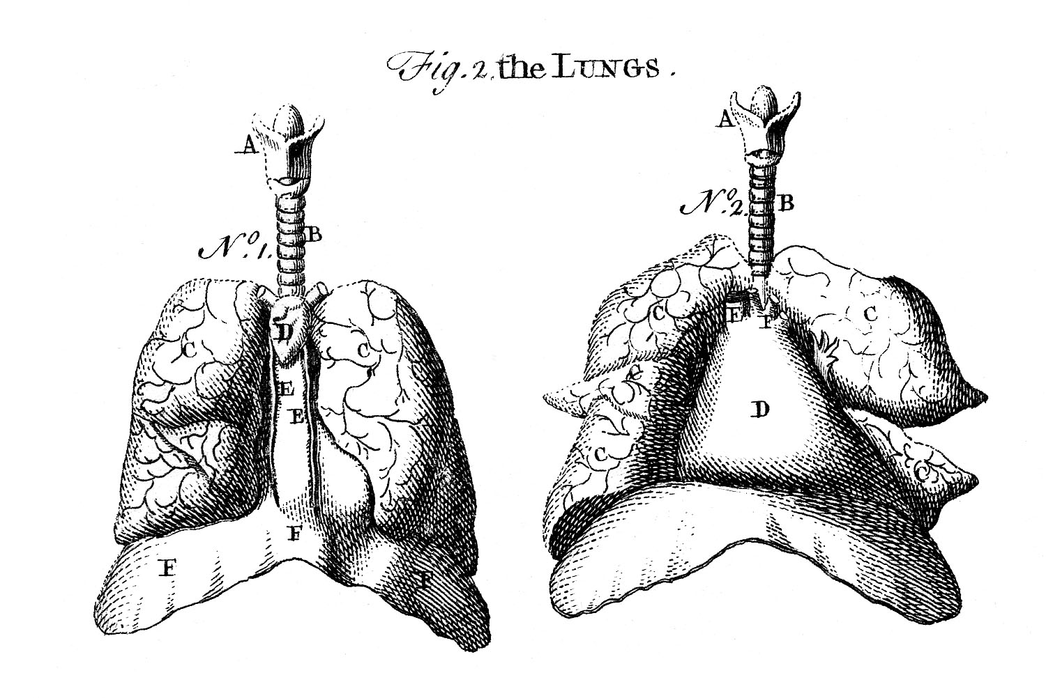
Early Anatomy Graphics - Diagram of Lungs - The Graphics Fairy
Lung - Wikipedia The lobes of the lungs can be seen, and the central root of the lung is also present. Right lung The right lung has both more lobes and segments than the left. It is divided into three lobes, an upper, middle, and a lower lobe by two fissures, one oblique and one horizontal. The upper, horizontal fissure, separates the upper from the middle lobe.

Dentistry and Medicine: Thorax,Lungs,Heart Anatomy and Physiology ...
How do Lungs Work Human Body Project for Kids Labeled Lungs. Now label the parts of the lung your child can grasp. How in-depth you go will really depend on the age of your student. Human Body Project. Insrt a straw into the completed paper bag. Gather the bag in around the straw, at the top of the sack and affix with packing tape.

Lung Structure | BioNinja
Download Anatomy Lung Images Download Anatomy Lung Images. Images. The lungs are the primary organs of the respiratory system in humans and many other animals including a few fish and some snails. There is little change in the topography of the lung from fetus to infant to adult. Anatomical Teaching Model Plastic Lung Model Lung Model With Larynx from .
Respiratory System Worksheet - WikiEducator
The Function, Anatomy, and Respiration of the Lungs The human body contains two lungs, of which one is positioned on the left side of the chest cavity and the other on the right side. The right lung is separated into three divisions or lobes, while the left lung contains two lobes. Each lung is surrounded by a two-layered membrane lining (pleura) that attaches the lungs to the chest cavity.
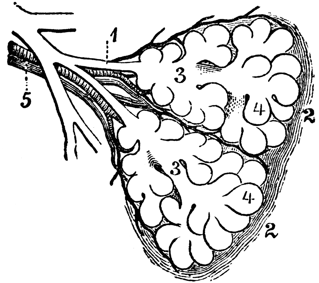
Diagram of the Two Primary Lobules of the Lung | ClipArt ETC
Hilum of the Lung: Definition, Anatomy, and Masses Anatomy of the Hilum Both the right and the left lung have a hilum which lies roughly midway down the lungs, and slightly towards the back (closer to the vertebrae than to the front of the chest). Each lung may be visualized as having an apex (the top), a base (the bottom), a root, and a hilum.
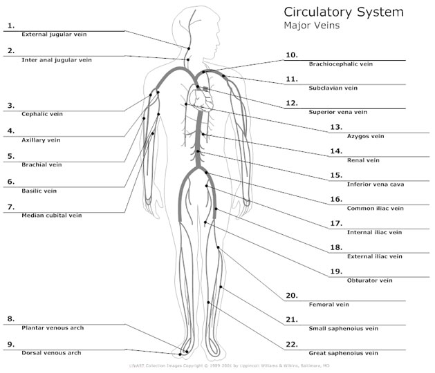
Circulatory System Diagram - Cardiovascular System and Blood ...
Histology Slides Identification from Different Organ Systems From the epithelial tissue, I should learn and identify the following histology slides with their identification points. #1. Simple squamous histology slide #2. Simple cuboidal epithelium histology slide #3. Simple columnar epithelium histology slide #4. Stratified squamous epithelial slide (both keratinized and non-keratinized) #5.
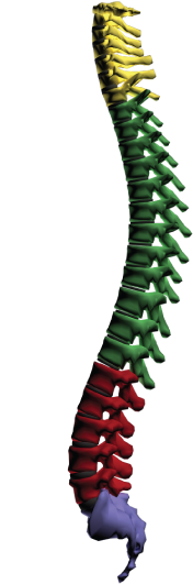
Spine Diagram - Active Chiropractic of Kansas City, MO
Chest anatomy illustrations - e-Anatomy - IMAIOS IASLC - Ganglionic areas: diagram of ganglionic areas numbered 1 to 14, used in clinical practice in thoracic oncology for lung cancer disease spread assessment. Anatomical structures of the respiratory system. 125 pulmonary anatomical structures were labeled.
Post a Comment for "43 diagram of the lungs with labels"