40 simple microscope diagram with labels
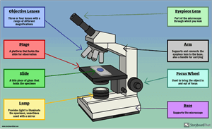
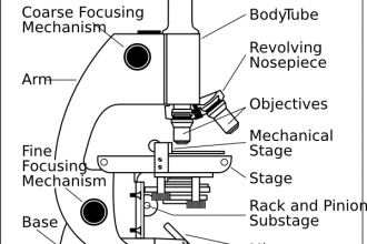
Parts of a microscope with functions and labeled diagram Structural parts of a microscope and their functions Figure created with biorender.com Figure: Diagram of parts of a microscope There are three structural parts of the microscope i.e. head, base, and arm. Head - This is also known as the body. It carries the optical parts in the upper part of the microscope. Base - It acts as microscopes support. Parts of the Microscope with Labeling (also Free Printouts) Parts of the Microscope with Labeling (also Free Printouts) A microscope is one of the invaluable tools in the laboratory setting. It is used to observe things that cannot be seen by the naked eye. Table of Contents 1. Eyepiece 2. Body tube/Head 3. Turret/Nose piece 4. Objective lenses 5. Knobs (fine and coarse) 6. Stage and stage clips 7. Aperture
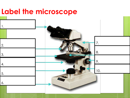
Parts of a Microscope Labeling Activity - Storyboard That Create a poster that labels the parts of a microscope and includes descriptions of what each part does. Click "Start Assignment". Use a landscape poster layout (large or small). Search for a diagram of a microscope. Using arrows and textables label each part of the microscope and describe its function. Copy This Storyboard* More options

Simple microscope diagram with labels
A Study of the Microscope and its Functions With a Labeled Diagram ... sciencestruck.com A Study of the Microscope and its Functions With a Labeled Diagram To better understand the structure and function of a microscope, we need to take a look at the labeled microscope diagrams of the compound and electron microscope. These diagrams clearly explain the functioning of the microscopes along with their respective parts. BYJUS Following are the parts of the simple microscope with their functions: Eyepiece: It is the lens that is used to study the samples and is placed at the top. It has a magnification of 10X to 15X. Base: This provides support to the microscope. Tube: This is used to connect the eyepiece to the objective lenses. Label the Microscope Diagram | Download Scientific Diagram the antibiogram of e. coli was investigated in different generations using eight antibiotic discs such as chloramphenicol (ch), streptomycin (s), gentamycin (g), ciprofloxacin (ci),...
Simple microscope diagram with labels. Draw a neat labeled diagram for the formation of an image in a simple ... A simple microscope is a convex lens where in the image is kept between the focus of the lens so that the image formed is virtual and magnified. While drawing make sure the symmetry is taken into consideration. Complete step by step answer: Here is the labeled diagram outlining a simple microscope. To draw the ray diagram, place the object ... Fluorescence Resonance Energy Transfer (FRET) Microscopy Presented in Figure 3 is a Jablonski diagram illustrating the coupled transitions involved between the donor emission and acceptor absorbance in fluorescence resonance energy transfer. Absorption and emission transitions are represented by straight vertical arrows (green and red, respectively), while vibrational relaxation is indicated by wavy ... Parts Of The Microscope Label Worksheets & Teaching Resources | TpT Check out this simple and clear 13 question worksheet that covers the parts of a microscope. Their is a visual at the top of the page that points to 13 parts. ... This simple 1-page worksheet has students label a diagram of a microscope using words from a word bank. Subjects: Basic Principles, Biology, General Science. Grades: 7 th - 12 th ... Labelled Diagram of Compound Microscope - Biology Discussion The below mentioned article provides a labelled diagram of compound microscope. Part # 1. The Stand: The stand is made up of a heavy foot which carries a curved inclinable limb or arm bearing the body tube. The foot is generally horse shoe-shaped structure (Fig. 2) which rests on table top or any other surface on which the microscope in kept.
Labeling the Parts of the Microscope | Microscope World Resources Labeling the Parts of the Microscope This activity has been designed for use in homes and schools. Each microscope layout (both blank and the version with answers) are available as PDF downloads. You can view a more in-depth review of each part of the microscope here. Download the Label the Parts of the Microscope PDF printable version here. Compound Microscope Parts - Labeled Diagram and their Functions - Rs ... There are three major structural parts of a compound microscope. The head includes the upper part of the microscope, which houses the most critical optical components, and the eyepiece tube of the microscope. The base acts as the foundation of microscopes and houses the illuminator. The arm connects between the base and the head parts. Compound Microscope Parts, Functions, and Labeled Diagram Compound Microscope Parts, Functions, and Labeled Diagram Parts of a Compound Microscope Each part of the compound microscope serves its own unique function, with each being important to the function of the scope as a whole. Parts of a Simple Microscope - Labeled (with diagrams) Parts of a Simple Microscope - Labeled (with diagrams) A simple microscope is a very first type of microscope ever created. It consists of simple parts and performs simple functions. Although there are now many advanced microscope types, some applications may still demand the use of a simple microscope.
Simple Microscope - Parts, Functions, Diagram and Labelling Simple Microscope - Parts, Functions, Diagram and Labelling A microscope is one of the commonly used equipment in a laboratory setting. A microscope is an optical instrument used to magnify an image of a tiny object; objects that are not visible to the human eyes. Table of Contents The common types of microscopes are: What is a Simple microscope? Plasmonics - an overview | ScienceDirect Topics The label-free feature of plasmonic imaging enables the non-invasive real-time observation of cellular processes but also raises challenges in identifying the cause of a local RI change. Thus, plasmonic imaging is applied mostly in simple systems where the imaging results can be interpreted by proper experimental design. Microscope labeled diagram - SlideShare Microscope labeled diagram 1. The Microscope Image courtesy of: Microscopehelp.com Basic rules to using the microscope 1. You should always carry a microscope with two hands, one on the arm and the other under the base. 2. You should always start on the lowest power objective lens and should always leave the microscope on the low power lens ... Microscope Types (with labeled diagrams) and Functions Simple microscope labeled diagram Simple microscope functions It is used in industrial applications like: Watchmakers to assemble watches Cloth industry to count the number of threads or fibers in a cloth Jewelers to examine the finer parts of jewelry Miniature artists to examine and build their work Also used to inspect finer details on products
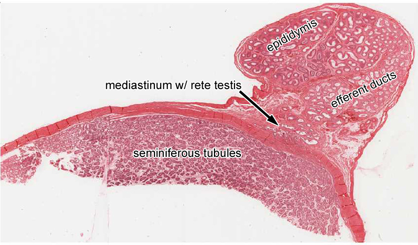
Male Reproductive System | histology
16 Parts of a Compound Microscope: Diagrams and Video Once you have an understanding of the parts of the microscope it will be much easier to navigate around and begin observing your specimen, which is the fun part! The 16 core parts of a compound microscope are: Head (Body) Arm. Base. Eyepiece. Eyepiece tube.
Parts of a Microscope Labeling Activity
Simple Microscope - Definition, Types, Working Principle & Formula A simple microscope consists of a convex lens of a short focal length. The below figure shows the ray diagram which subsequently forms the image of an object (or we can say a source of light). (Image will be Updated soon) F is the focal length of the lens. An object is placed between the focal length and the centre of the curvature.
Microscope Labelled Diagram Gcse - Micropedia
Microscope Drawing And Label - Painting Valley label microscope diagram compound parts light labeling functions microscopic blank labeled biology microscopy labelled beautiful Compound Microscope ... 496x600 35 0 Parts Of A Compound ... 500x469 27 0 Microscopic Drawing ... 1024x1024 21 4 Download The Diagram... 547x579 17 0 Microscope Labeling ... 270x350 17 0 Microscope Labeling ...

Biology Notes – ElimuTanzania
Label the microscope — Science Learning Hub All microscopes share features in common. In this interactive, you can label the different parts of a microscope. Use this with the Microscope parts activity to help students identify and label the main parts of a microscope and then describe their functions. Drag and drop the text labels onto the microscope diagram.
Respiratory System Clip Art at Clker.com - vector clip art online, royalty free & public domain
Microscope Labeling - The Biology Corner 1) Start with scanning (the shortest objective) and only use the COARSE knob . Once it is focused… 2) Switch to low power (medium) and only use the COARSE knob . You may need to recenter your slide. Once it is focused.. 3) Switch to high power (long objective).

All Saints Online: Diagram for Labelling: Microscope
PDF Parts of a Microscope Printables - Homeschool Creations Label the parts of the microscope. You can use the word bank below to fill in the blanks or cut and paste the words at the bottom. Microscope Created by Jolanthe @ HomeschoolCreations.net. Parts of a eyepiece arm stageclips nosepiece focusing knobs illuminator stage objective lenses

Microscope Unlabelled Diagram - Micropedia
Optical Sensor - an overview | ScienceDirect Topics A far-field, epifluorescence microscope system for single tube spectroscopy was first proposed by Weisman and co-workers, and its schematic diagram is presented in Fig. 10.11. 23, 27 Visible images of sample morphology can be viewed through the eyepiece of the microscope or through a CCD camera. The samples are photo-excited by lasers, and ...
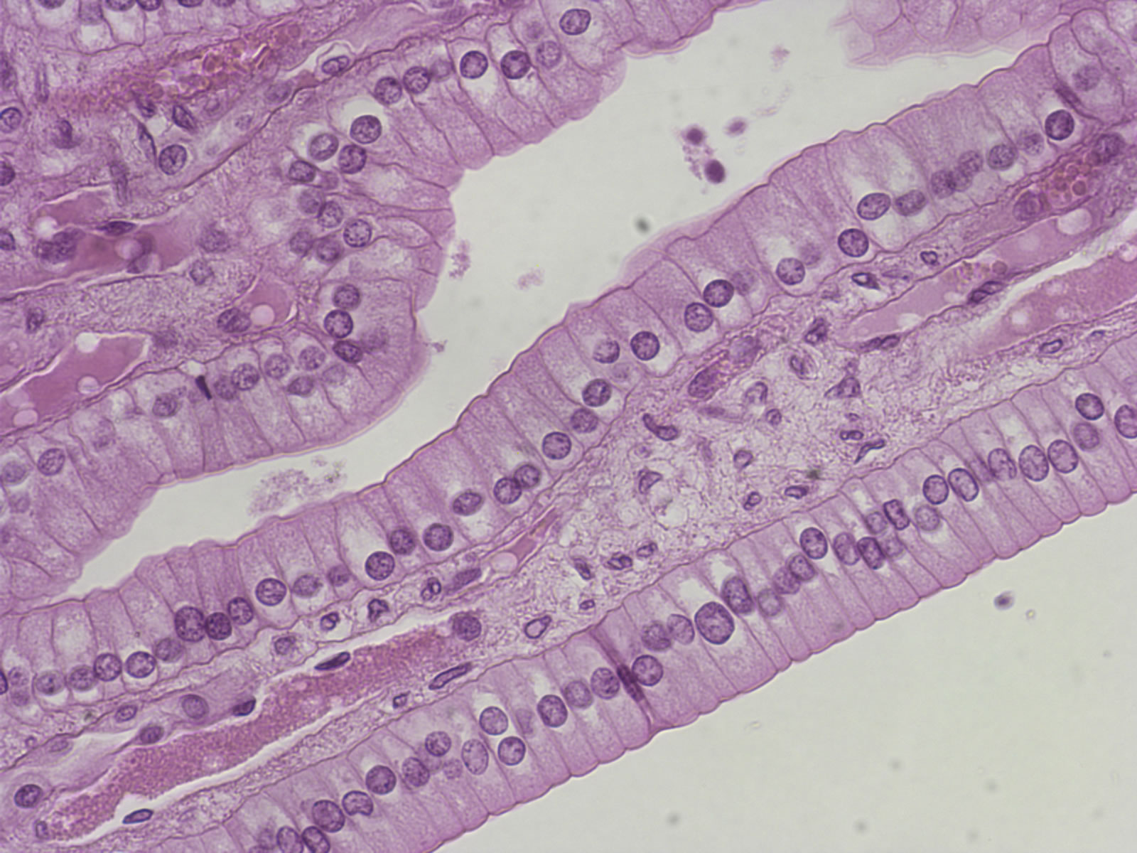
Flashcards Table on Tissues For Anatomy
Microscope, Microscope Parts, Labeled Diagram, and Functions Microscope, Microscope Parts, Labeled Diagram, and Functions What is Microscope? A microscope is a laboratory instrument used to examine objects that are too small to be seen by the naked eye. It is derived from Ancient Greek words and composed of mikrós, "small" and skopeîn,"to look" or "see".
NCERT Exemplar Class 9 Science Solutions Chapter 5 - The fundamental Unit of Life | Download ...
Microscope Labeling - The Biology Corner Students label the parts of the microscope in this photo of a basic laboratory light microscope. Can be used for practice or as a quiz. ... The type of microscope used in most science classes is the _____ microscope. 18. You should carry the microscope by the _____ and the _____. 19. The objectives are attached to what part of the microscope ...

Easy labeled diagram of Microscope - YouTube
Interactive Bacteria Cell Model - CELLS alive Ribosomes: Ribosomes give the cytoplasm of bacteria a granular appearance in electron micrographs.Though smaller than the ribosomes in eukaryotic cells, these inclusions have a similar function in translating the genetic message in messenger RNA into the production of peptide sequences (proteins).

ปักพินโดย ð“¥ð“®ð“𓪠ð“. ใน Biology | Science biology Secondary school science และ Science diagrams
Simple Microscope - Diagram (Parts labelled), Principle, Formula and Uses Parts of a Simple Microscope A simple microscope consists of Optical parts Mechanical parts Labeled Diagram of simple microscope parts Optical parts The optical parts of a simple microscope include Lens Mirror Eyepiece Lens A simple microscope uses biconvex lens to magnify the image of a specimen under focus.
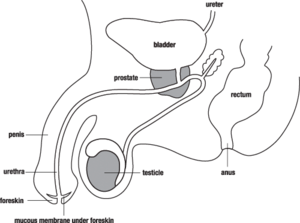
Male Reproductive System | Free Images at Clker.com - vector clip art online, royalty free ...
Labeling Microscope Worksheet | Teaching Resources File previews. docx, 300.56 KB. A straightforward worksheet in which students are required to identify the parts of a basic microscope. Tes classic free licence.
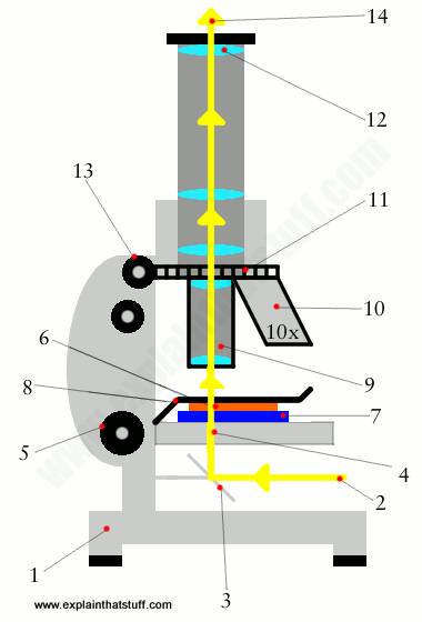
How does a microscope work? - Explain that Stuff
Microscope Poster - Diagram with Labels | Teach Starter A poster containing a diagram with labels showing the key parts of a microscope. In Science it is important that students know how to use a variety of tools when conducting scientific experiments and inquiry. This poster focuses on the microscope and highlights its key parts. There are two print options available for this poster:
Microscope With Labels clip art : Biological Science Picture Directory – Pulpbits.net
Compound Microscope- Definition, Labeled Diagram, Principle, Parts, Uses The optical microscope often referred to as the light microscope, is a type of microscope that uses visible light and a system of lenses to magnify images of small subjects. There are two basic types of optical microscopes: Simple microscopes. Compound microscopes. The term "compound" in compound microscopes refers to the microscope having ...

All Saints Online: Diagram for Labelling: Microscope
Anatomy Chart - How to Make Medical Drawings and Illustrations Pathologic anatomy focuses on how diseases affect and change the human body. Histology studies microscopic anatomy such as tissues and cells visible only under a microscope. Anatomy charts serve two main purposes: education in the form of anatomy worksheets and presentation in the form of simple healthcare illustrations.

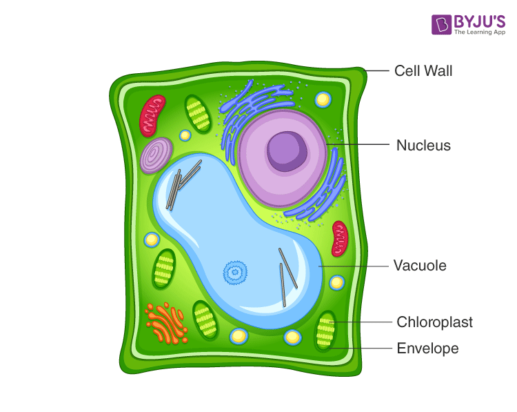
The Parts Of An Animal Cell | Science Trends
Answered: Learning through Art: Water Molecules… | bartleby Transcribed Image Text:

Introductory Biology
Label the Microscope Diagram | Download Scientific Diagram the antibiogram of e. coli was investigated in different generations using eight antibiotic discs such as chloramphenicol (ch), streptomycin (s), gentamycin (g), ciprofloxacin (ci),...
Post a Comment for "40 simple microscope diagram with labels"