40 eye diagram and labels
Eye anatomy: A closer look at the parts of the eye Eye anatomy: A closer look at the parts of the eye. By Liz Segre. When surveyed about the five senses — sight, hearing, taste, smell and touch — people consistently report that their eyesight is the mode of perception they value (and fear losing) most. Despite this, many people don't have a good understanding of the anatomy of the eye, how ... Eye Diagram Teaching Resources | Teachers Pay Teachers The Human Eye Overview Reading Comprehension and Diagram Worksheet. by. Teaching to the Middle. 63. $1.50. Zip. This passage briefly describes the human eye (900-1000 Lexile). 14 questions (matching and multiple choice) assess students' understanding. Students label a diagram of 6 parts of the eye. I've included a color and BW version, as well ...
Labeled Eye Diagram - Pinterest Human Eye Anatomy Diagram · Eye Anatomy L'Optique Optometry Rochester Hills, MI 248.656.5055 · The Auditory System | in Chapter 04: ...
Eye diagram and labels
Eye Diagram With Labels and detailed description - BYJUS A brief description of the eye along with a well-labelled diagram is given below for reference. Well-Labelled Diagram of Eye The anterior chamber of the eye is the space between the cornea and the iris and is filled with a lubricating fluid, aqueous humour. The vascular layer of the eye, known as the choroid contains the connective tissue. Eye Anatomy: Parts of the Eye and How We See Behind the anterior chamber is the eye's iris (the colored part of the eye) and the dark hole in the middle called the pupil. Muscles in the iris dilate (widen) or constrict (narrow) the pupil to control the amount of light reaching the back of the eye. Directly behind the pupil sits the lens. The lens focuses light toward the back of the eye. Eye Diagram Stock Photos, Pictures & Royalty-Free Images - iStock Browse 4,092 eye diagram stock photos and images available, or search for human eye diagram or eye diagram vector to find more great stock photos and pictures. Anatomy of human eye and descriptions. Components of human eye. Illustration about Anatomy and Physiology. Human eye anatomy.
Eye diagram and labels. Labeled Eye Diagram | Science Trends The human eye is composed of many different parts that work together to interpret the world around us. What you want to interpret as a major part of the human eye is somewhat up to the individual, but in general there are seven parts of the human eye: the cornea, the pupil, the iris, the lens, the vitreous humor, the retina, and the sclera. Let's take a closer look at each of these ... The Eyes (Human Anatomy): Diagram, Optic Nerve, Iris, Cornea ... - WebMD Iris: the colored part. Cornea: a clear dome over the iris. Pupil: the black circular opening in the iris that lets light in. Sclera: the white of your eye. Conjunctiva: a thin layer of tissue ... Anatomy of the Human Eye - News-Medical Retina is the innermost layer of the eyeball structure. Retinal membrane can be imagined as the wall on which the images are projected. The light passing ... eye diagram labeled Eye anatomy. eye diagram labeled. The brain - structure and function - Cancer Information - Macmillan we have 8 Images about The brain - structure and function - Cancer Information - Macmillan like Eye Anatomy | Eye Care | Eye Health | Prince Frederick MD | Annapolis MD, Basic Eye Anatomy Quiz and also The brain - structure and function ...
Diagram of the Eye Side View No Labels Illustration - Twinkl Sep 18, 2019 - Diagram of the Eye Side View No Labels,Eye,Science,Human Body,Biology,Diagram,Human,Pupil,Sense,Organ,Sclera,Cornea,Iris,Lens,Retina,Optic ... Vision and Eye Diagram: How We See - AARP Cones are responsible for producing the visual sharpness of the eye — seeing road signs when driving, fine print when reading or recognizing facial details like the color of someone's eyes — as well as color vision. "Most of these cones are concentrated in a very specific area in the center of retina, called the macula," says Haugsdal. Human eye diagram, Biology diagrams, Eye structure Human eye diagram and functions How to draw human eye diagram step by step Eye diagram for kids Diagram of human eye with labelling Human eye diagram easy ... Eye Diagram: Label Quiz - PurposeGames.com Tournaments (37) AI Stream The more you play, the more accurate suggestions for you. Cities by Landmarks 11p Image Quiz. Cities of Midwestern US 32p Image Quiz. I spy on... 26p Image Quiz. The Western States 11p Image Quiz. Highscores (6 registered players) Member. Score.
Labelling the eye - Science Learning Hub In this interactive, you can label parts of the human eye. Use your mouse or finger to hover over a box to highlight the part to be named. Drag and drop the text labels onto the boxes next to the eye diagram. If you want to redo an answer, click on the box and the answer will go back to the top so you can move it to another box. The Eye - diagram to label | Teaching Resources File previews. pdf, 2.94 MB. Diagram of eye with key words to use in labelling it. Tes classic free licence. Eye Diagram Stock Illustrations - 6,878 Eye Diagram ... - Dreamstime How the eye works medical scheme poster, elegant and minimal vector illustration, eye - brain labeled structure diagram. Stylized and artistic medical design. Diagram of cataract in human eye. Illustration. Human Eye Anatomy Diagram. Eye anatomy 3d diagram infographics layout showing human eyes muscles in side view with labeling vector illustration Anatomy of the Eye Diagrams for Coloring/Labeling, with Reference and ... This printable contains 13 clear and simple cross sectional diagrams of the human eye. They photocopy well and are great for use as a labeling and coloring exercise for your students. The core eye anatomy diagram, designed as the labeling exercise, has a fully colored and labeled reference chart to go with it.
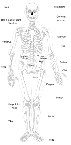
Labeled Skeleton Clip Art at Clker.com - vector clip art online, royalty free & public domain
PDF Parts of the Eye - National Eye Institute | National Eye Institute Macula: The macula is the small, sensitive area of the retina that gives central vision. It is located in the center of the retina. Optic nerve: The optic nerve is the largest sensory nerve of the eye. It carries impulses for sight from the retina to the brain. Pupil: The pupil is the opening at the center of the iris.
Horseshoe Crab Anatomy
Labelled Diagram of Human Eye, Explanation and Function - VEDANTU Labeled Diagram of Human Eye The eyes of all mammals consist of a non-image-forming photosensitive ganglion within the retina which receives light, adjusts the dimensions of the pupil, regulates the availability of melatonin hormones, and also entertains the body clock.
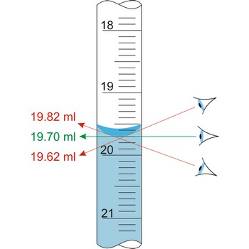
Meniscus @ Chemistry Dictionary & Glossary
Labeled Eye Diagram | Eye anatomy diagram, Eye anatomy, Diagram of the eye This Article is the detailed account of all the major organs that are categorized under the nine regions in the abdominal cavity 1) Stomach 2) Intestines a) Small Intestine Duodenum Jejunum Ileum b) Large Intestine Ceacum Colon (Ascending, Transverse and Descending) Rectum Anal Canal 3) Liver 4) Gall bladder 5) Pancreas 6) Spleen 7) Kidneys ...
Post a Comment for "40 eye diagram and labels"